

The 44th Congress of Polish Physical Society

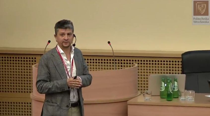

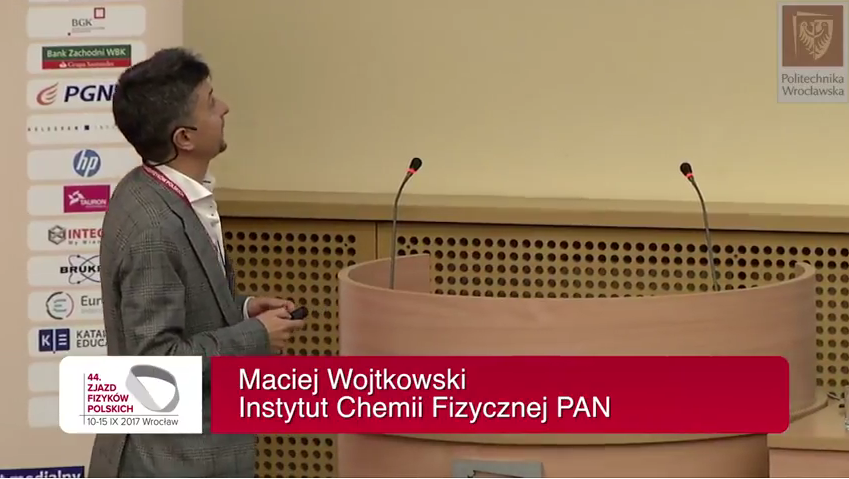
Professor Maciej Wojtkowski participated in the 44th Congress of Polish Physical Society in Wroclaw, with a plenary lecture entitled "Journey from organs to cells: In vivo imaging by spatio-temporal optical coherence techniques".
The Congress of Polish Physical Society is the oldest Polish physics conference, with its history reaching back to the Inaugural Meeting held in Warsaw in 1923 and since then it has been organized in various cities in Poland by local branches of the Polish Physical Society. The 44th Congress took place at the Congress Centre of Wroclaw University of Science and Technology and was co-organized by the Wrocław University of Science and Technology, the University of Wrocław and the Institute of Low Temperature and Structural Research. This year it was held under the Honorary Patronage of both Ministry of Science and Higher Education of Poland and the Mayor of Wroclaw. The lectures selected for the Congress showcased the most important recent research advances in physics, with emphasis on achievements that involved Polish contribution. One of the specialized parallel afternoon sessions was devoted to optics, as a branch of physics especially strongly developed in the Department of Optics and Photonics of the Wroclaw University of Technology.
The plenary lecture, gave by Maciej Wojtkowski on 11.09.2017, has been focused on the most appealing and still unsolved problems in biological and medical imaging: the possibility of non-invasive visualization of tissue in vivo with an accuracy of microscopic examination.
Abstract of the lecture:
One of the most appealing and still unsolved problems in biological and medical imaging is the possibility of non-invasive visualization of tissue in vivo with an accuracy of microscopic examination. It is especially emphasized nowadays in era of novel microscopic techniques, which have ability for optical depth sectioning without need of processing samples.
The main physical limitation of the in vivo microscopic imaging is associated with light scattering introduced by irregular and often discontinuous distribution of refractive index. Scattering of light limits the number of ballistic photons delivered to and received from the sample. As a consequence, the contrast of reconstructed images is compromised dramatically by increased noise. Another side effects of uneven distribution of the refractive index are significant deformations of images. Additionally, in case of coherent illumination with laser light there is a disturbing presence of so-called speckles – strong fluctuations of intensity caused by interference of mixed transverse modes of the laser beam. Speckle noise also degrades system resolution and reduces image quality. Adding all of these effects results in severe loss of imaging information.
In our work we try to solve these fundamental physical limitations by developing new optical coherence imaging techniques, which utilize spatio-temporally partially coherent light with access to intensity and phase of detected radiation. In our research activity we focus on developing new optical methods that enable to image biological objects in vivo and in minimally invasive way. We went long way covering significant spectrum of various sizes of objects – from organ-size scale up to internal structure of a single cell.
The Congress served as a great platform for exchanging knowledge and experience between various groups of physicists-researchers, which allows to broaden the perspective and start new collaborations.
A presentation was recorded and published by the organizer at the conference YouTube channel
Dates
September 10-15, 2017
Location
Wroclaw, Poland
Organizers
Polish Physical Society,
Wroclaw University of Technology,
Wroclaw University,
Institute of Low Temperature and Structure Research PAS
More information about the event .