

Visit to the University of California Irvine
Jadwiga Milkiewicz, a member of the POB group, visited the University of California Irvine (US). The main goal of this visit was to build a high-resolution two-photon excited fluorescence microscope for retinal imaging in-vivo.
Imaging autofluorescence from the eye fundus is already known and used by ophthalmologists to diagnose a health condition of the retinal pigment epithelium (RPE). One of the good sides of this technique is that it can be done without an additional contrast agent, as the naturally occurring pigments of the retina can emit fluorescence after absorption of the light. The energy of the light needed for this process corresponds to the range of UV and short wavelengths of the visible light. In practice, such devices use bright blue light, which can excite some of these pigments.
The approach in the team of professor Wojtkowski is slightly different than commonly used. Red light (or even infrared) is used to stimulate the fluorescence of retinal pigments. This is possible if the molecule simultaneously absorbs two consecutive photons; therefore, as a light source, we need to use short pulse lasers. In this phenomenon fluorescence occurs at a shorter wavelength than the wavelength of the exciting light itself. Using far red light has its advantages, as it is not very disturbing (or even unpleasant) for the patient, as the intense blue light, and it is less scattered by the front of the eye.
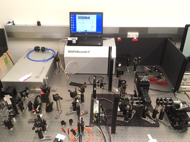
The laboratory which Jadwiga Milkiewicz visited already has a commercial version of such a two-photon excited fluorescence microscope, which can be used for detailed ophthalmic research of small mammals. This microscope is very large and expensive. Our goal was to build one with similar performance, but much smaller and cheaper, which could be later adapted to human and easily used by medical doctors. The main construction of the small microscope has already been built during this internship.
Visitor
Jadwiga Milkiewicz
Dates
28 Jan.-5 Apr. 2020
Location
University of California Irvine, USA
Figures
Fig. 1 Construction of small two-photon excited fluorescence microscope.
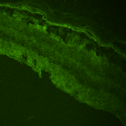
Fig. 2 Two-photon excited fluorescence image of mice retinal section.