

Working visit to Optical Biomedical Imaging Group Institute of Physics, Nicolaus Copernicus University.
The visit to Optical Biomedical Imaging Group Institute of Physics, Nicolaus Copernicus University was to have the deep analysis of the in-vivo imaging of the global mouse brain ischemia (GI) using Bessel beam optical coherence microscopy. This method allows to monitor changes in brain structure with extra control of blood flow during the process of artery occlusion. The results showed the capability and sensitivity of OCM system with Bessel beam to analyze brain plasticity after severe injury within a period of 8 days.
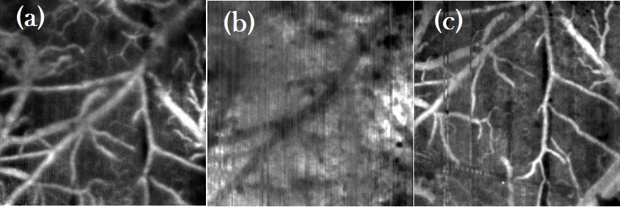
This visit has helped to analyze the angiographic and structural B-scan maps along with microscopic images of the mouse brain gives the GI information and allow to visually monitor the induced structural change in brain before global ischemic stroke, at the time of the stroke and up to at least 8 days after the stroke. It provides the detail understanding of the underlying pathway and shows evidence that this particular mouse model had survived the stroke. The deeper analysis of the structural changes after stroke is yet to be made. This experiment sheds some light for the better understanding of the global ischemia of the brain and may lead to findings saving many life’s and reducing the disability with improving the quality of life. It may also be a convenient tool for testing new drugs against ischemia-induced neurodegeneration.
Visitor
Mounika Rapolu
Dates
January 9-10, 2017
Location
Torun, Nicolaus Copernicus University