

Classification of biological micro-objects using optical coherence tomography: in silico study
BIOMEDICAL OPTICS EXPRESS 2017 Vol. 8, No. 8 | 1 Aug | 3606-3626
PAWEŁ OSSOWSKI, MACIEJ WOJTKOWSKI AND PETER RT MUNRO
Abstract:
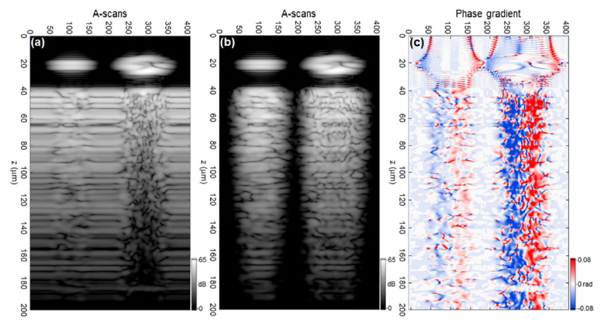
We report on the development of a technique for differentiating between biological micro-objects using a rigorous, full-wave model of OCT image formation. We model an existing experimental prototype which uses OCT to interrogate a microfluidic chip containing the blood cells. A full-wave model is required since the technique uses light back-scattered by a scattering substrate, rather than by the cells directly. The light back-scattered by the substrate is perturbed upon propagation through the cells, which flow between the substrate and imaging system’s objective lens. We present the key elements of the 3D, Maxwell equation-based computational model, the key findings of the computational study and a comparison with experimental results.